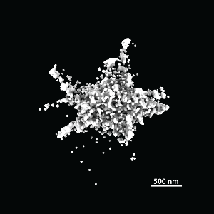
Endogenous ASC speck imaged in 3D by dSTORM
The ASC speck is a central component of the NLRP3 inflammasome. | © iScience
A central component of the human immune system, the NLRP3 inflammasome plays an important role in fighting off infections. However, its chronic activation is also implicated in a variety of common diseases, such as Alzheimer’s, Parkinson’s, multiple sclerosis, atherosclerosis, gout, and type II diabetes. The NLRP3 inflammasome occurs primarily in specialized immune cells in the blood and elsewhere. It is a dense complex in which several proteins interact with each other. A key protein in this complex is known by the abbreviation ASC. In non-activated immune cells, it is distributed homogeneously throughout the cell. If the NLRP3 inflammasome is activated, all of the ASC protein present in the cell aggregates in the inflammasome complex. Under an ordinary fluorescence microscope, the ASC protein, once labeled, appears as a single, bright, nearly round spot. Due to the small size and high density of this ASC speck, scientists have been unable to elucidate details of its structure inside cells. Different models were proposed in the scientific literature but a comprehensive understanding was missing.
An international team of researchers, including the research groups of LMU professors Don Lamb, Ralf Jungmann, and Veit Hornung, has now visualized the 3D structure of the ASC speck inside cells using various fluorescence microscopy methods. Recently published in the journal iScience, their study shows that the ASC speck has an amorphous structure with a dense core from which filaments reach out into the periphery. To be able to fully label and image the structure, the researchers had to combine two different approaches. They labeled the less dense periphery of the ASC speck with antibodies and the dense interior with nanobodies.